Pediatric MRI: Indications, Safety and Procedure Details
MRI is a safe, non-radiation diagnostic method suitable for children of any age, including newborns. Learn about indications and cases where anesthesia is required.
Read more →Magnetic Resonance Imaging (MRI) at Brigid Medical Center utilizes modern 1.5 Tesla technology to produce exceptionally detailed images of internal structures without ionizing radiation. This advanced imaging method provides unsurpassed soft-tissue contrast, allowing for the precise diagnosis of neurological, musculoskeletal, abdominal, and vascular diseases with the highest level of patient safety.
Zero ionizing radiation — completely safe for children, pregnant women, and repeat examinations.
Excellent visualization of the brain, spinal cord, muscles, ligaments, and internal organs.
High-quality images in axial, sagittal, coronal, and oblique planes without repositioning the patient.
Diffusion, perfusion, spectroscopic, and functional MRI for comprehensive neurological assessment.
Reduced examination time through parallel imaging and accelerated scan sequences.
Detection of lesions as small as millimeters and early pathological changes before symptoms appear.
Magnetic Resonance Imaging (MRI) is a non-invasive diagnostic method that uses powerful magnetic fields and radio waves to create detailed images of organs and tissues. Unlike CT scans, MRI does not use ionizing radiation, making it one of the safest imaging modalities available. The 1.5 Tesla magnet strength provides the optimal balance between image quality, scan speed, and patient comfort.
Comprehensive safety screening for metal implants, devices, or foreign bodies. Changing into specialized clinical attire and removing all metallic objects. Planning contrast administration if necessary.
Comfortable positioning on the scanning table. Headphones are provided for communication and scanner noise reduction. An emergency call button is placed within the patient's reach.
The table moves into the magnet bore. A series of images are captured according to the protocol for the area being studied. Patients are guided through breath-holding instructions if required.
Immediate review of captured images by the radiologist or technologist to ensure diagnostic quality. Additional sequences may be performed if necessary to clarify specific clinical questions.
Assistance exiting the scanning table. Post-contrast monitoring if applicable. Preliminary findings discussion. A report from a certified radiologist is typically provided within 24–48 hours.
Magnetic Resonance Imaging provides unsurpassed visualization for accurate diagnosis and effective treatment planning.
Contact Brigid Medical Center to schedule your MRI or consult with our radiologists regarding the optimal imaging method for your diagnostic needs.
Brigid Medical Center — Leading MRI Diagnostics in Kharkiv.
1.5 Tesla Technology · Radiation-Free Imaging · Exceptional Contrast · Expert Interpretation
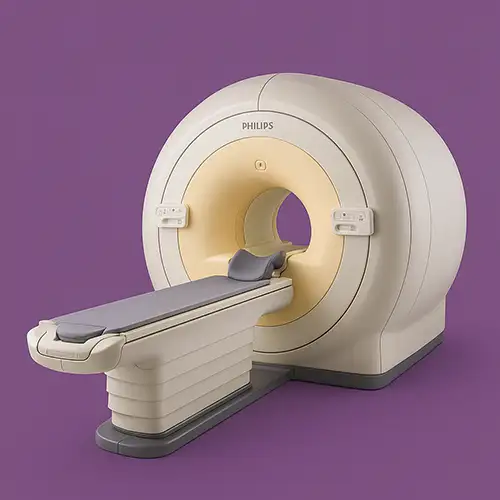
A high-field MRI system delivering excellent image quality, fast acquisition, and exceptional patient comfort. Suitable for comprehensive diagnostics across all clinical applications.


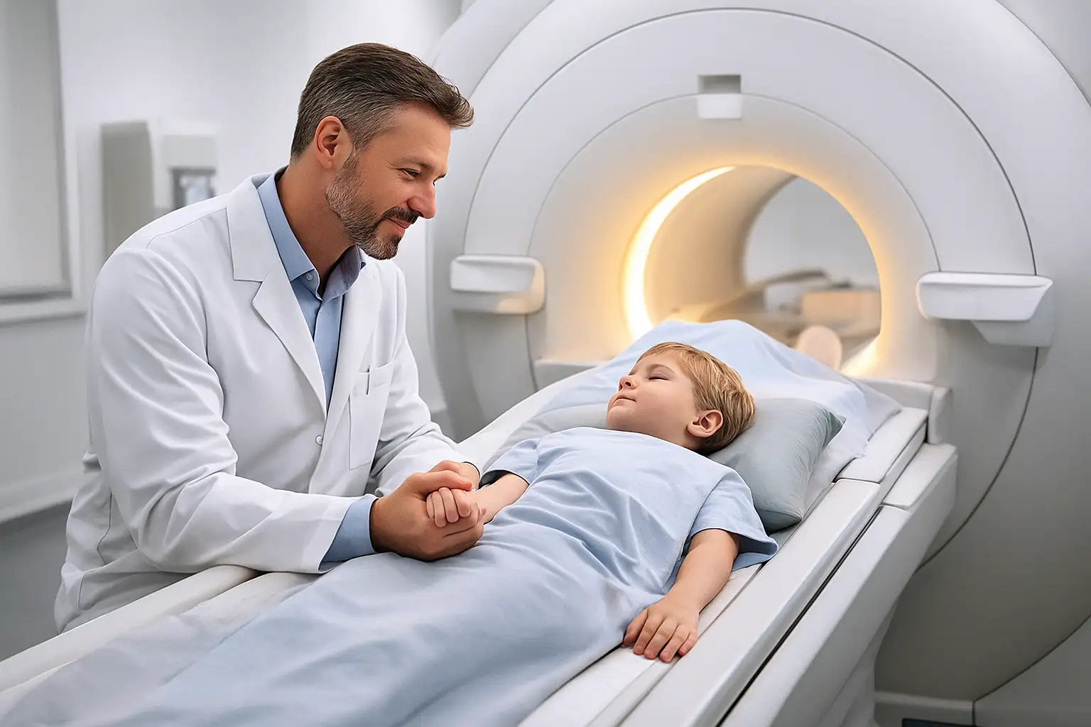
MRI is a safe, non-radiation diagnostic method suitable for children of any age, including newborns. Learn about indications and cases where anesthesia is required.
Read more →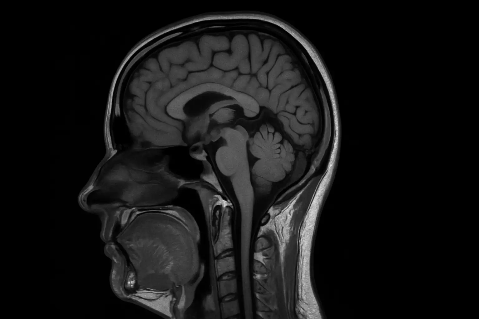
Brain MRI and MRA are complementary diagnostic methods. Learn the differences, indications for each test, and how to prepare for high-accuracy imaging.
Read more →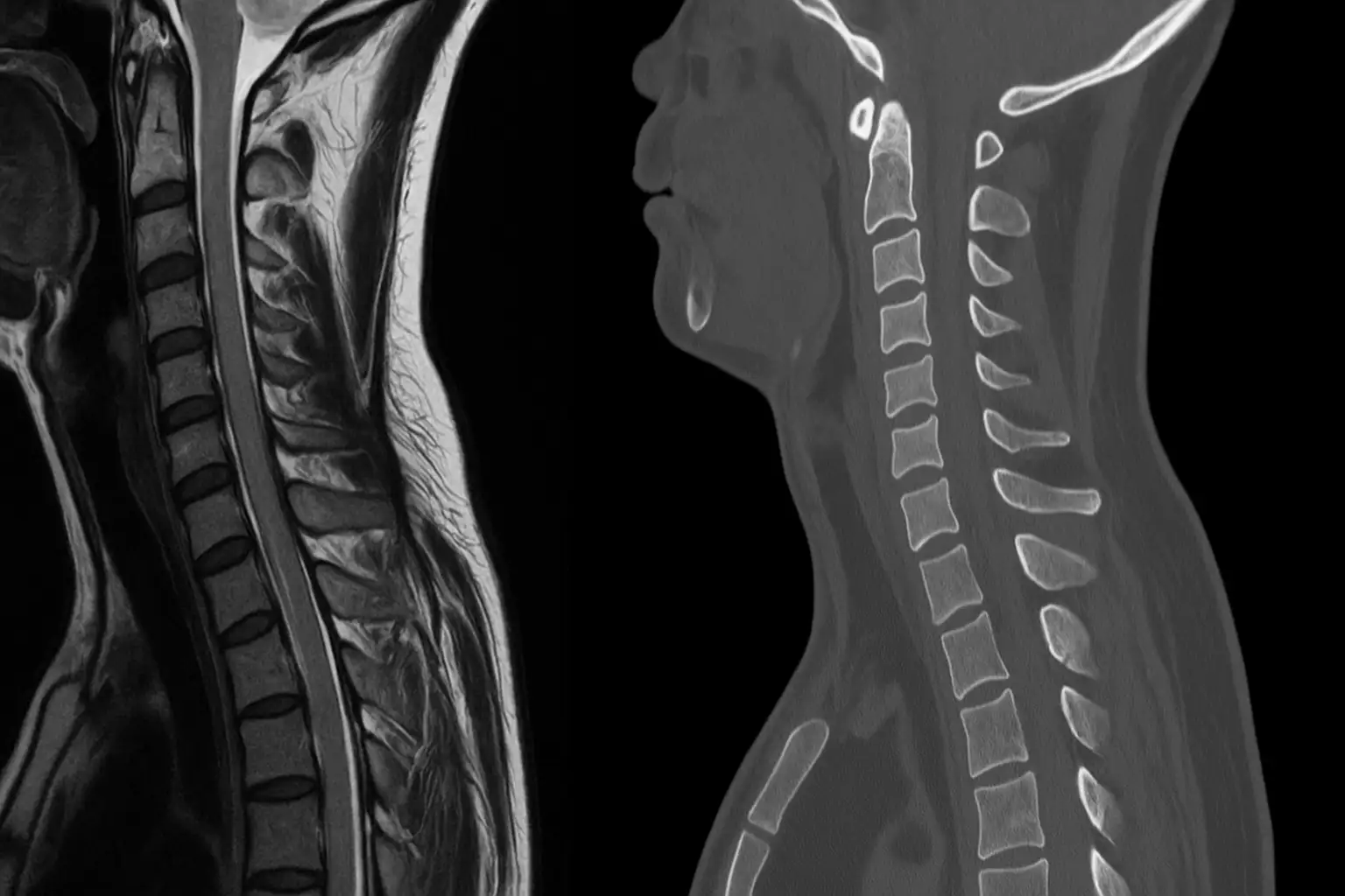
CT and MRI are two advanced imaging methods for the spine. Learn the key differences, indications for each test, and how to choose the right diagnostic path.
Read more →
Pelvic MRI is a highly accurate, non-invasive imaging method used to diagnose gynecologic disorders, inflammation, and tumors at early stages. The procedure is safe and provides detailed visualization of the pelvic organs.
Read more →
Online booking is preliminary. We will contact you to clarify details and confirm the appointment.
Your personal data is fully protected and not shared with third parties. Contact information is needed only for feedback and appointment scheduling.