Ultrasound Diagnostics in Kharkiv — Advanced Imaging Technologies
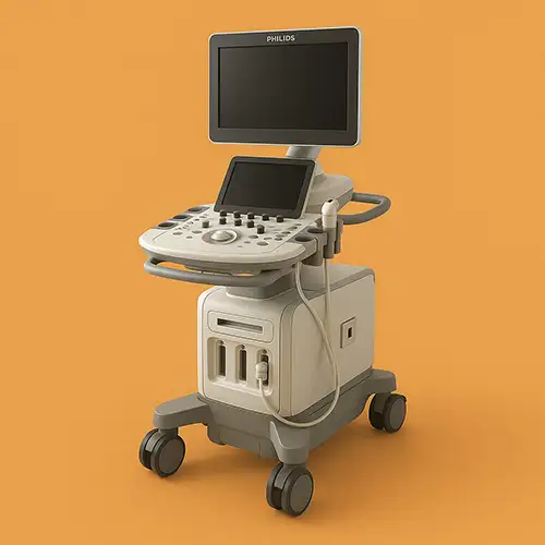
Ultrasound Diagnostics (Ultrasound) at Brigid Medical Center is performed using the state-of-the-art Philips EPIQ Elite ultrasound system, which provides exceptional image quality, advanced diagnostic capabilities, and maximum patient comfort. This premium diagnostic platform allows for the detailed visualization of organs, blood vessels, and tissues with unsurpassed clarity, ensuring accurate diagnosis and effective treatment planning.
Key Advantages of the Philips EPIQ Elite Ultrasound System
🔬
Crystal Clear Imaging
nSIGHT technology provides resolution up to 5 times better than conventional ultrasound systems.
🛡️
100% Safe and Non-Invasive
Zero radiation exposure — safe for pregnant women, children, and all patient groups.
📊
Advanced Quantification
Automated measurements and AI-driven analysis for objective diagnostic data.
💓
Cardiological Excellence
Premium cardiac imaging featuring 4D Echo and strain analysis.
🤰
4D Fetal Visualization
Realistic 4D fetal imaging with HDlive technology for detailed prenatal assessment.
⚡
Shear Wave Elastography
Measurement of tissue stiffness for non-invasive liver fibrosis staging without biopsy.
What is Modern Ultrasound Diagnostics?
Ultrasound diagnostics is a non-invasive imaging method that uses high-frequency sound waves to create real-time images of internal organs and structures. Unlike other imaging modalities, ultrasound involves no ionizing radiation, making it the safest diagnostic method for pregnancy monitoring, pediatric patients, and repeat examinations.
100%
Safe — zero radiation risk for any patient group
0.5 mm
Spatial resolution to detect the smallest pathological changes
15–45
Minutes — average duration of an examination
Real-Time
Imaging allows for the dynamic assessment of organ function
When is an Ultrasound Recommended?
Abdominal and Pelvic Diagnostics:
- Liver Assessment — fatty liver disease, cirrhosis, tumors, cysts.
- Gallbladder Study — stones, polyps, inflammation.
- Pancreatic Evaluation — pancreatitis, tumors, cystic lesions.
- Kidneys and Urinary Tract — stones, tumors, structural anomalies.
- Spleen Examination — size, structure, focal lesions.
- Abdominal Aorta — screening for aneurysms and atherosclerosis.
Obstetrics and Gynecology:
- Pregnancy Monitoring — fetal development, anatomical screening, growth assessment.
- Gynecological Exam — evaluation of the uterus, ovaries, and fallopian tubes.
- Fertility Assessment — ovarian reserve, follicle tracking (folliculometry).
- Early Pregnancy — viability and detection of ectopic pregnancy.
- Pelvic Floor Evaluation — prolapse and urinary incontinence.
- Breast Ultrasound — supplemental diagnostic for mammography and dense breast tissue.
Cardiovascular and Vascular Diagnostics:
- Echocardiography — assessment of cardiac structure and function.
- Doppler Ultrasound — analysis of blood flow velocity and direction.
- Carotid Arteries — screening for atherosclerosis and stenosis.
- Peripheral Vessels — DVT screening, arterial insufficiency, varicose veins.
- Abdominal Vessels — portal vein, hepatic veins, renal arteries.
Musculoskeletal and Superficial Structures:
- Joint Studies — arthritis, bursitis, tendon injuries.
- Muscle Evaluation — tears, hematomas, myositis.
- Thyroid Gland — nodules, goiter, inflammation.
- Lymph Nodes — size, structure, malignancy assessment.
- Soft Tissue Masses — characterization of lipomas, cysts, tumors.
The Ultrasound Examination Process
1
Preparation and Consultation
Review of medical history and explanation of the procedure. Specific preparation instructions depending on the exam type (e.g., fasting for abdominal scans or a full bladder for pelvic scans).
2
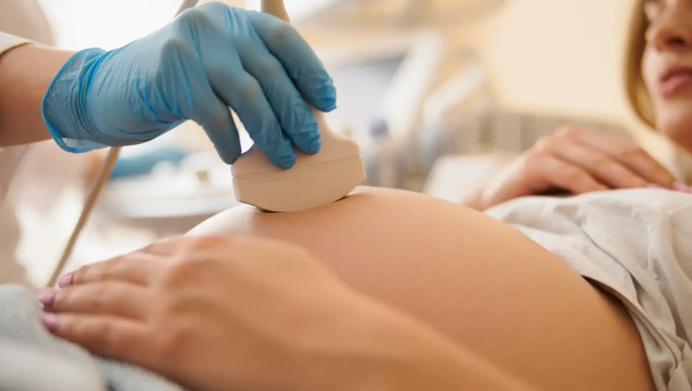
Positioning and Setup
Comfortable positioning on the examination table; application of warm ultrasound gel to ensure optimal transducer contact and image quality. Room temperature is adjusted for patient comfort.
3
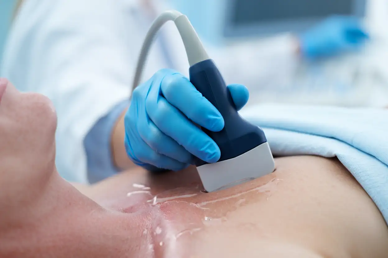
Scanning Process
Systematic scanning of target organs using appropriate transducers and settings. Real-time imaging allows for the dynamic assessment of organ function and blood flow.
4
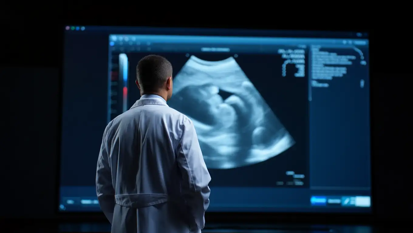
Image Analysis and Measurement
Detailed evaluation of anatomical structures, automated measurements, and Doppler spectrum analysis. Diagnostic images and video loops are saved for documentation.
5
Results and Explanation
Immediate discussion of preliminary findings and answering patient questions. A formal report from a certified sonologist is provided within 24 hours.
Patient Safety and Comfort
Ultrasound is the safest imaging method with numerous advantages:
- Zero Radiation — completely safe for repeat examinations.
- Safety in Pregnancy — FDA-approved for all trimesters.
- Pediatric Safety — ideal for children of all ages.
- Non-Invasive — no needles, incisions, or contrast agents required.
- Painless — a comfortable examination with minimal preparation.
- Immediate Results — real-time visualization allows for instant assessment.
Schedule Your Ultrasound Examination
Accurate and timely diagnosis detects conditions at their earliest stages. Our Philips EPIQ Elite system ensures premium quality in a patient-centered environment.
Contact Brigid Medical Center to book an ultrasound or consult with our sonologists regarding the most appropriate imaging approach for your needs.
Brigid Medical Center — Advanced Ultrasound Diagnostics in Kharkiv.
Philips EPIQ Elite · Crystal Clear Imaging · 100% Safe · 4D Technology · Expert Interpretation