Low-Field Joint MRI 0.35T in Kharkiv — Esaote O-scan Technology with Open Design
Low-field MRI using the Esaote O-scan device at the Brigid Medical Center provides high-precision imaging of joints and limbs using innovative open-type technology. This specialized system offers exceptional visualization of ligaments, tendons, cartilage, menisci, and bone structures with maximum patient comfort, eliminating claustrophobia issues while ensuring diagnostic quality comparable to high-field systems for joint examinations.
Key Benefits of Esaote O-scan Low-Field MRI
High Resolution
Specialized surface coils and optimized protocols for detailed visualization of joint structures
Fully Open Design
No tunnel — only the limb is scanned, the patient sits comfortably, ideal for claustrophobic patients
Fast Scanning
Complete joint examination in 30-40 minutes with rapid image reconstruction
Enhanced Safety
Lower magnetic field strength allows for examining patients with certain implants and metal constructions
Joint Specialization
Technology specifically developed for musculoskeletal imaging with excellent soft tissue contrast
Quiet Operation
Significantly lower noise level compared to conventional high-field MRI systems
What is Esaote O-scan Low-Field MRI?
The Esaote O-scan represents a breakthrough in specialized extremity MRI technology, utilizing a 0.35 Tesla magnetic field specifically optimized for joint visualization. Unlike traditional closed-bore MRI systems, this open design focuses exclusively on peripheral joints, ensuring exceptional diagnostic quality for orthopedic, rheumatological, and sports medicine applications with maximum comfort and accessibility for patients.
When is Low-Field MRI Recommended?
Symptoms and conditions requiring joint MRI:
- Persistent joint pain of unknown origin lasting more than 2-3 weeks
- Post-traumatic injuries after falls, sports injuries, or direct impacts
- Swelling and inflammation of joints that do not improve with conservative treatment
- Mechanical symptoms such as blocking, locking, or joint instability
- Limited mobility or stiffness in the joints
- Sensation of instability or recurrent joint dislocations
- Suspected sports injuries in athletes and active individuals
- Post-operative evaluation after arthroscopy or joint surgeries
- Monitoring treatment effectiveness for arthritis or degenerative diseases
- Pre-operative planning for orthopedic surgical interventions
Specific joint diseases we diagnose:
- Ligament damage: ACL, PCL, MCL, LCL tears and strains
- Meniscal pathology: tears, degeneration, discoid meniscus
- Tendon diseases: tendinitis, tendinosis, partial and complete tears
- Cartilage damage: chondromalacia, osteochondral defects, arthritis
- Bone marrow edema: bone bruises, stress reactions, osteonecrosis
- Inflammatory conditions: synovitis, bursitis, rheumatoid arthritis
- Degenerative changes: osteoarthritis, joint space narrowing
- Sports injuries: rotator cuff tears, labral tears, impingement syndromes
- Fractures: occult fractures, stress fractures, osteochondral fractures
- Soft tissue masses: cysts, ganglia, tumors in the joint area
Patient groups who benefit the most:
- Claustrophobic patients who cannot tolerate traditional MRI
- Patients with certain implants not compatible with high-field MRI
- Pediatric patients requiring joint imaging in comfortable conditions
- Elderly patients who need assistance during the examination
- Overweight patients exceeding the limits of traditional scanners
- Athletes requiring rapid diagnosis
Which Joints We Examine on Esaote O-scan
Knee Joint
- Evaluation of ACL, PCL, MCL, LCL ligaments
- Evaluation of medial and lateral menisci
- Analysis of articular cartilage and bone marrow
- Patellofemoral joint and retinaculum
- Detection of effusion and synovitis
Ankle Joint and Foot
- Lateral and medial ligament complexes
- Achilles tendon and plantar fascia
- Tarsal tunnel and sinus tarsi
- Osteochondral lesions of the talus
- Detection of stress fractures in foot bones
Hand and Wrist Joint
- TFCC (triangular fibrocartilage complex) evaluation
- Carpal tunnel and Guyon's canal evaluation
- Ligament injuries and instability
- Tendon pathology and tenosynovitis
- Evaluation of small joint arthritis
What to Expect During an MRI Examination
Preparation and Screening
Medical history review and screening for contraindications. Removal of metal objects and changing clothes if necessary. Detailed explanation of the procedure and positioning.
Comfortable Positioning
Positioning while sitting in a comfortable chair with the limb placed in a specialized coil. Adjustments for optimal comfort and image quality. No need to enter a closed tunnel.
Scanning Process
Quiet scanning process with minimal noise. Ability to communicate with the technologist throughout the procedure. Multiple sequences to capture different tissue characteristics. Average duration 15-30 minutes.
Image Processing
Fast image reconstruction using advanced algorithms. Initial quality check by the MRI technologist. Transfer of images to a radiologist for detailed interpretation.
Results and Consultation
Expert analysis by a radiologist and report preparation. Results available on the day of the study. Discussion of results with the referring physician or direct patient consultation.
Technical Capabilities of Esaote O-scan MRI
Our Esaote O-scan system is equipped with advanced technology for superior joint imaging:
- 0.35 Tesla Magnetic Field: Optimized specifically for peripheral joint imaging
- Specialized Surface Coils: High-sensitivity coils designed for each joint type
- Open Design: 270-degree open access without tunnel restrictions
- Advanced Software: Specialized protocols and sequences for the musculoskeletal system
- Fast Imaging: Short scanning time while maintaining diagnostic quality
- Low Noise Level: Quiet operation enhancing patient comfort
- Weight Capacity: Accommodates patients up to 200 kg
- Compatibility: Broader compatibility with implants and medical devices
Schedule Your Low-Field MRI Examination
Early and accurate diagnosis of joint diseases is essential for effective treatment and recovery. Whether you are experiencing sports injuries, chronic joint pain, or post-traumatic symptoms, our Esaote O-scan provides detailed visualization of joint structures without the discomfort of traditional MRI. The open design makes this technology accessible to patients who previously could not undergo MRI examinations due to claustrophobia or body size limitations.
Contact us today to schedule your low-field MRI examination. Experience comfortable, high-quality joint imaging with fast results and expert interpretation.
Brigid Medical Center — Modern Low-Field Joint MRI in Kharkiv.
Esaote O-scan Technology · Open Design · Expert Radiologists · Results on the Day of Study · Comfortable Experience
Our Equipment
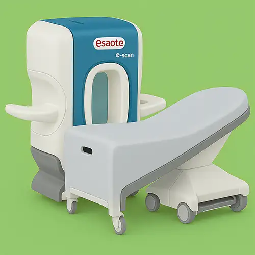
Esaote O-Scan MRI 0,35 Tl
A modern, high-quality MRI system designed specifically for joint and limb imaging, without the need to enter a full-body tunnel. Perfect for claustrophobic patients and children.
- Open design — no feeling of tight or enclosed space
- Accurate imaging of knees, ankles, feet, wrists, elbows, and other joints
- Low noise level for a calm and comfortable exam
- Short, patient-friendly examination time
- Ideal for athletes and post-injury evaluations
Our Doctors

Palchyk Serhiy Mykhailovych

Panasenko Vladislav Alexandrovich

Kutsyn Vladyslav Mykolayovych

Baliasna Viktoria Valeriyivna

Online booking is preliminary. We will contact you to clarify details and confirm the appointment.
Your personal data is fully protected and not shared with third parties. Contact information is needed only for feedback and appointment scheduling.