Reducing Radiation Dose on CT: How IMR Technology Ensures Safety
Is it possible to reduce the radiation dose on CT without losing image quality? Yes, it is already a standard in modern diagnostics. At Brigid Medical Center, we use advanced Philips CT scanners equipped with Iterative Model Reconstruction (IMR) technology to significantly reduce exposure while improving detail.
What is IMR (Iterative Model Reconstruction)?
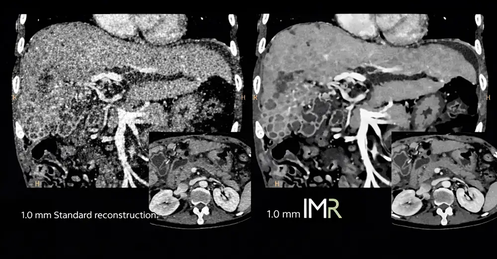
IMR is an intelligent algorithm that "cleans up" medical images from digital noise and artifacts. Think of it like a professional photo editor: it removes graininess, enhances fine details, and produces a sharp image even from minimal raw data.
Key Benefits for the Patient
- Up to 80% Lower Dose: Makes repeated examinations and preventative screenings much safer;
- Superior Detail: Improves detection of low-contrast structures (soft tissues, small lesions) by 43–80%;
- Noise-Free Imaging: Provides "quiet" images, crucial in oncology and neurology where every millimeter counts;
- Cardiac Precision: Compensates for heart motion during high-speed coronary angiography (CTA).
Who Benefits Most?
- Children and young adults with radiosensitive tissues;
- Oncology patients requiring regular treatment monitoring;
- Screening patients (e.g., lung cancer screening);
- Patients with complex conditions where maximum resolution is required to see small metastases.
Investing in Philips CT with IMR reflects our commitment to the ALARA principle — using the lowest dose possible while maintaining uncompromised diagnostic quality.